Mitochondria
Outer Membrane
The outer membrane contains proteins known as porins, which allow movement of ions into and out of the mitochondrion.
Enzymes involved in the elongation of fatty acids and the oxidation of adrenaline can also be found on the outer membrane.
The space within the inner membrane of the mitochondrion is known as the matrix, which contains the enzymes of the Krebs (TCA) and fatty acid cycles, alongside DNA, RNA, ribosomes and calcium granules.
The inner membrane contains a variety of enzymes. It contains ATP synthase which generates ATP in the matrix, and transport proteins that regulate the movement of metabolites into and out of the matrix.
The inner membrane is arranged into cristae in order to increase the surface area available for energy production via oxidative phosphorylation.
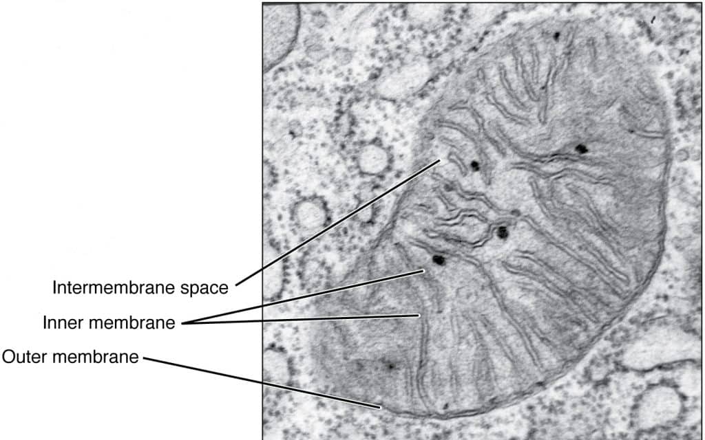
Fig 1 – Electron micrograph of a mitochondrion
By Rice University [CC BY 4.0], via openstax CNX
Function
Mitochondria also produce chemicals that your body needs for other purposes, break down waste products so they’re less harmful, and recycle some of those waste products to save energy.
Mitochondria also have a special role in making cells die (apoptosis). This may sound strange, but it is vital for the processes of growth and development. Sometimes cells don’t die when they should, and start to grow uncontrollably. This is how a tumour starts to grow, so you shouldn’t be surprised that mitochondria play an important part in cancer and are seen as targets for anti-cancer drugs.
To produce all of that energy, mitochondria require oxygen. Mitochondria effectively burn your food in a carefully controlled way to produce that chemical energy by a process called “oxidative phosphorylation”.
During a heart attack, or a stroke, the blood stops delivering oxygen to the heart and brain. These two organs do a lot of work and need a lot of energy. Without oxygen, the mitochondria stop working, and the cells in the brain or heart are damaged or even die. Perversely, if the oxygen does return, then the mitochondria get overwhelmed and produce a lot of “free radicals”. These are very reactive chemicals which cause a lot of additional damage - called “Reperfusion injury”.
The mitochondrion is the site of ATP synthesis for the cell. The number of mitochondria found in a cell are therefore a good indicator of the cell’s rate of metabolic activity; cells which are very metabolically active, such as hepatocytes, will have many mitochondria.
Mitochondria also have a role to help maintain the intracellular environment. They:
- Store caspases responsible for triggering apoptosis.
- Are able to transiently store calcium contributing to calcium homeostasis.
In brown adipose tissue mitochondria have an alternative function of heat production using the electron transport chain.
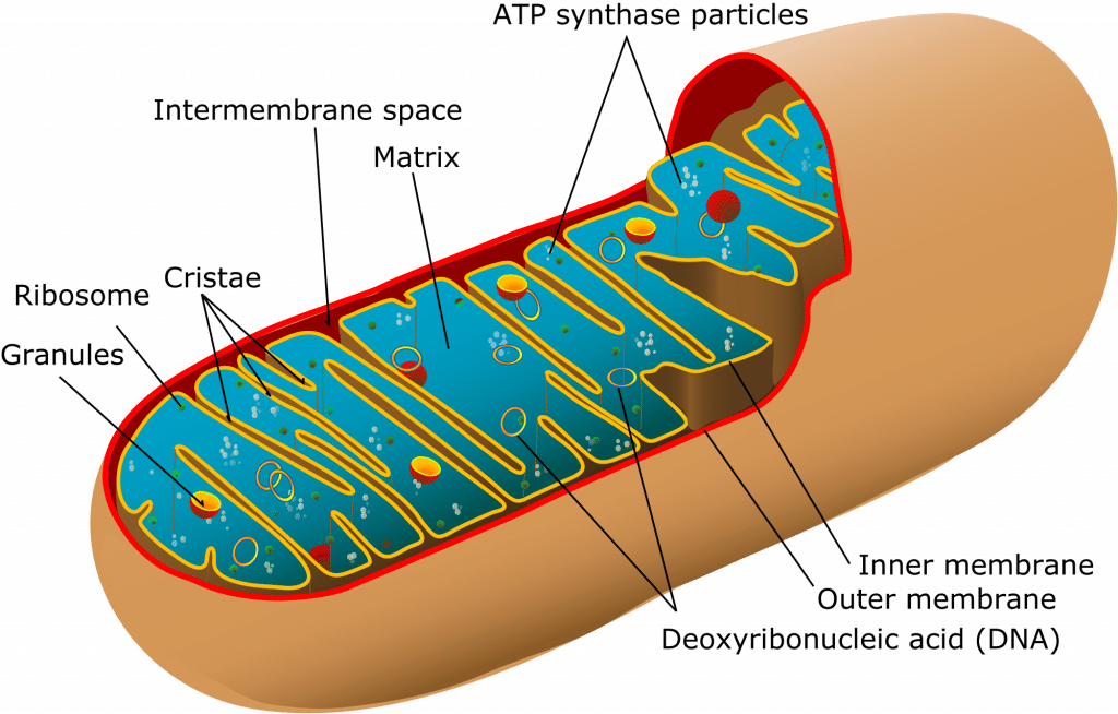
Fig 2 – A mitochondrion with its main features
By Mariana Ruiz Villarreal LadyofHats [Public Domain], via Wikimedia Commons
DNA and Inheritance
Mitochondria replicate their DNA by a process called binary fission and can use this to make multiple copies in one mitochondrion.
Their DNA has maternal lineage which means their DNA is passed from mother to child with little change.
Clinical Relevance - Leber’s Hereditary Optic Neuropathy
As a result of how crucial mitochondria are to the survival of the cell, mitochondrial disorders are rare.
Some general features of mitochondrial disease include exercise intolerance, myopathy and muscle weakness.
The most common mitochondrial disease is Leber’s Hereditary Optic Neuropathy (LHON) which affects the optic nerve causing blurring of central vision and loss of colour vision and carries a risk of developing blindness.
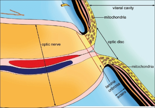
Fig 3 – Mitochondria in the optic nerve
Abu-Amero, KK. [CC BY 2.0], via OPENi
A transmission electron micrograph of a mitochondrion in a chick embryo cell. (Prof. R Bellairs/Wellcome Images)
Each mitochondrion has an outer membrane, an intermembrane space, an inner membrane, which is folded to form shelves (cristae), and a central matrix space. The enzyme complexes responsible for oxidative phosphorylation are lined up on the cristae.

The darker spots in the image are ribosomes occupying the mitochondrial matrix. These are involved in protein synthesis.
A single mitochondrion has two membranes, with the inner membrane folded into inward projections, known as cristae.
It is here where final stage of aerobic respiration takes place, with an electron transport chain generating energy in the form of adenosine triphosphate (ATP).
The more metabolically active a cell, the more mitochondria it will have, and with more cristae per mitochondrion.
Inside the organelle, the fluid around the cristae is known as the mitochondrial matrix. This hosts the earlier link reaction and Krebs cycle stages of aerobic respiration.

Components involved in oxidative phosphorylation in mitochondria and their origins. As enzyme complexes I through IV convert 2-carbon metabolic fragments to CO2 and H2O, protons (H+) are pumped into the intermembrane space. The protons diffuse back to the matrix space via complex V, ATP synthase (AS), in which ADP is converted to ATP. The enzyme complexes are made up of subunits coded by mitochondrial DNA (mDNA) and nuclear DNA (nDNA), and the figures document the contribution of each DNA to the complexes.
Mitochondirial Genome
Mitochondria have their own genome. Human mitochondrial DNA is a double-stranded circular molecule containing approximately 16,500 base pairs (compared with over a billion in nuclear DNA). It codes for 13 protein subunits that are associated with proteins encoded by nuclear genes to form four enzyme complexes plus two ribosomal and 22 transfer RNAs that are needed for protein production by the intramitochondrial ribosomes. Mitochondria have an ineffective DNA repair system; the mutation rate for mitochondrial DNA is over 10 times the rate for nuclear DNA. A large number of relatively rare diseases have been traced to mutations in mitochondrial DNA. These include disorders of tissues with high metabolic rates in which energy production is defective as a result of abnormalities in the production of ATP, as well as other disorders
++++++++++++++++
Classically referred to as the ‘powerhouse of the cell’, they are the site of the majority of ATP synthesis and are therefore exceptionally important to function both microscopically and macroscopically.
recycling centres, and assassins...
Mitochindria turn sugars, fats and proteins that we eat, into forms of chemical energy that the body can use to carry on living.
Oxidative Phosphorylation
Mitochondria provide cells with the ability to form the energy-rich compound ATP by oxidative phosphorylation under aerobic conditions.1
This process is mediated by the respiratory electron transport chain (ETC) multiprotein enzyme complexes I–V and the two electron carriers, coenzyme Q (CoQ) and cytochrome c.
Given the centrality of oxidative phosphorylation to the normal activities of almost all cells, it is not surprising that mitochondrial dysfunction can affect almost any organ system.
Apoptosis
Mitochondria make a major contribution to apoptosis (programmed cell death) and additional cell type–specific functions.
The efficiency of the mitochondrial ETC in ATP production is a major determinant of overall body energy balance and thermogenesis.
In addition, mitochondria are the predominant source of reactive oxygen species (ROS), whose rate of production also relates to the coupling of ATP production to oxygen consumption.
Additionally, a variety of potentially toxic molecules are sequestered within the mitochondria, including large stores of calcium as well as key regulators of apoptosis such as Cytochrome C.